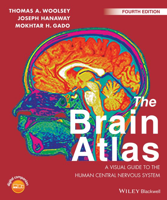
The Brain Atlas
Praise for earlier editions:
"... an essential requirement for the library of any individual who works in the field... if you buy only one atlas, this is the one to buy."Journal of Neurosurgery
"... an excellent tool for understanding the central nervous system. It is a good companion at every level of training and for health care professionals."Archives of Neurology
"... The Brain Atlas presents neuroanatomy in an intuitive yet comprehensive manner, which makes it an excellent reference book and an outstanding learning text for anyone trying to master the intricate layout of the brain and its related structures."Trends in Neuroscience
The Brain Atlas: A Visual Guide to the Human Central Nervous System demonstrates brain anatomy in cross-referenced detail to be used to explain the anatomical basis for neurologic diseases and has been extensively revised and updated for a fourth edition making it the best available visual guide to human neuroanatomy.
The human brain is one of the most amazing consequences of evolution. The Brain Atlas correlates human brains, anatomical slices of brains, as well as MRI and MRA scans showing brain structure in three different planes. This text includes clearly labeled details of all parts of the brain, color illustrations and diagrams of nerve pathways, and easy navigation with color coding and brain-section markers on each page.
The Brain Atlas:
- Shows brain structures and the interneuronal connections that clarify human neuroanatomy and relate to function and disease without overwhelming users with detail and/or oversimplifying the brain
- Uses direct labeling system around the specimen including an alphabetical list of terms for each image
- Contains approximately 350 high quality images showing the brain in incredible detail
- Features unrivaled treatment of brain pathways, with colored lines that clearly trace pathways on actual brain slices detailed in the anatomical section of the book
- Shows systematic correlation of magnetic resonance images side-by-side with corresponding brain slices
- Includes blood supply maps to show arteries and veins of the CNS in detail, and separate vascular territory maps directly correlated to each whole brain slice
- Comes with free access to Wiley companion digital edition accessible on any device, allowing the reader to explore interactive learning functionality on figures, make notes, bookmark, and follow cross references
This book is ideal for undergraduate and graduate medical students, and for trainee neurologists, neurosurgeons, psychiatrists, psychologists, neuroscientists, and neurobiologists. Its easy to understand layout makes it a must have resource for anyone who wants a deep and clear working knowledge of brain anatomy.
The Brain Atlas: A Visual Guide to the Human Central Nervous System integrates modern neuroscience with clinical practice and is now significantly revised and updated for a Fourth Edition. The book's five sections cover: Background Information, The Brain and Its Blood Vessels, Brain Slices, Histological Sections, and Pathways. These are depicted in over 350 high quality intricate figures making it the best available visual guide to human neuroanatomy.
| Auteur | | Ta Woolsey |
| Taal | | Engels |
| Type | | Paperback |
| Categorie | | Wetenschap & Natuur |


