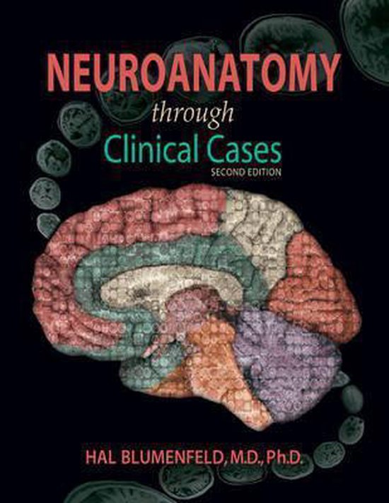
Neuroanatomy Through Clinical Cases
The goal of this book is to provide a treatment of neuroanatomy that ts com prehensive, yet enables students to focus on the most important “take-home messages” for each topic. This goal is motivated by the recognition that, while access to detailed information is often useful in mastering neuroanatomy, certain selected pieces of information carry the most clinical relevance, or are most important for exam review.
General Outline
The first four chapters of the book contain introductory material that will be especìally useful to students who have little previous clinical background. Chapter 1 is an introduction to the standard format commonly used for presenting clinical cases, including an outline of the medical history, physical examination, neuroanatomical localization, and differential diagnosis. Chapter 2 is a brief overview of neuroanatomy which includes definitions and descriptions of basic structures that will be studied in greater detail in later chapters. Chapter 3 builds on this knowledge by describing the neurologic examination. It includes a summary of the structures and pathways tested in each part of the exam, which is essential for localizing the lesions presented in the clinical cases throughout the remainder of the book. Much of the material in this chapter is also covered on the neuroexam.com website described below, which provides video demonstrations for each part of the exam. For readers who are unfamiliar with neuroimaging techniques, Chapter 4 contains a concise introduction to CT, MRI, and other imaging methods. This chapter also includes a Neuroradiological Atlas showing normal CT, MRI, and angiographic images of the brain. Chapters 5-19 cover the major neuroanatomical systems and present relevant clinical cases.
Chapters 5-19
Chapters 5-19 have a common structure. An “Anatomical and Clinical Review” at the beginning of the chapters presents relevant neuroanatomical structures and pathways, and generously sized, carefully labeled color illustrations are used to vividly depict spatial relationships. The first part of each chapter also includes numbered sections called “Key Clinical Concept,” or “KCC,” which cover common disorders of the system being discussed.
CLINICAL CASES The second part of each chapter is a “Clinical Cases” section that deserbes patients seen by the author and colleagues, each presented in a numbered color box. Full-length cases include complete findings from the neurologie evamination, while “Minicases” have a briefer format. Each case begins with à narrative of how the patient’s symptoms developed and what deticits were found on neurologie evamination. For example, one patient in Chapter 10 suddenly developed weakness in the right hand and lost the abilitv to speak. Another, in Chapter 14, experienced double vision and lapsed inte a coma, Important symptoms and signs are indicated in boldface type. The reader ìs then challenged through a series of questions to deduce the neuroanatomical location of the patient’s lesion and the eventual diagnosis.
A discussion follows each case, beginning with a summary of the key symptoms and signs. Answers to the questions are provided which refer to anatomical and clinical material presented in the first half of the chapter that is demonstrated bv the case. Continual improvements in imaging technology have allowed us to make clear and detailed radiographs of the nervous system in vvo, and one of the most exciting features of the book is the inclusion of largetormat, labeled CT, MRI, or other scans that show the lesion for each patient, and serve as a central tool for teaching neuroanatomy. These images reveal, with striking clarity, both the lesion’s location and the anatomy of the system being studied. In addition, these radiographs help the reader develop skill in interpreting the kinds of diagnostic images employed on the wards. The neuramaging studies for each case are provided in special boxes at least one page turn away from the case questions, so the answers to the questions are not ‘given away” by the imaging (see below).
The clinical course is also provided for each patient, and includes a discusson of how the patient was managed, and what outcome followed. Thus, by the end of each case, students learn the relevant material by application and diagnostic sleuthing rather than by rote memorization.
Special Features for Focused Study and Review
Since one of the goals of this book is to enable students to either read the ma
terial in depth, or to distill it down to the most clinically relevant points or to
material most commonly covered on the national boards or other examinations, several special features have been included to expedite focused study and review:
e Boldface type is used rather differently than in most texts. In addition to identifying the text for all important topics and definitions, boldface is also used to facilitate rapid or focused reading.
e Review Exercises appear in the margins throughout the text, highlighting the most important anatomical concepts in each chapter, and providing practice exam questions.
* Helpful mnemonics are provided throughout the text, and these are flagged in the margins by a special icon (shown at left) showing a section of the hippocampus (a structure important in memory formation).
* A Brief Anatomical Study Guide appears at the end of each chapter, which summarizes the most important neuroanatomical material, and refers to the appropriate figures and tables needed for focused exam review.
e The Neuroradiological Atlas in Chapter 4 also provides a useful review of neuroanatomical structures in three-dimensional space, and can be used for reference and comparison to lesions seen in clinical cases.
| Auteur | | Hal Blumenfeld |
| Taal | | Engels |
| Type | | Paperback |
| Categorie | | Wetenschap & Natuur |
